Why Choose Cortical Fixation Implant Systems?

Choosing the right implant platform requires more than comparing product features. Cortical fixation implant systems are designed to engage dense cortical bone, supporting stable fixation in carefully selected clinical situations. Their value depends on anatomy, bone quality, loading demands, and the treatment objective.

For clinicians, the decision should begin with patient assessment and high-quality imaging. A narrow screw corridor, fragile bone, or altered anatomy may change the preferred approach. Experienced surgeons also consider insertion technique, construct alignment, implant compatibility, and the possibility of revision. Small details matter, including tactile feedback during placement and the accuracy of postoperative imaging.

Evidence should guide confidence. Peer-reviewed studies, manufacturer testing, documented material specifications, and appropriate regulatory clearance provide useful reference points. However, laboratory strength does not automatically predict every patient outcome. Clinical judgment remains essential. It should not be replaced by marketing language.

A well-designed system may offer efficient fixation, controlled load transfer, and procedural flexibility. Yet these advantages can involve trade-offs, such as technical sensitivity, cost, or limited suitability for unusual anatomy. Not every patient needs the same solution. That matters.

This overview examines why clinicians may choose these systems, how their design supports fixation, and where caution remains necessary. It also recognizes an uncomfortable truth: even strong evidence cannot remove surgical uncertainty. Careful planning, verified technique, and long-term follow-up help turn implant performance into dependable patient care.
Why Choose Cortical Fixation Implant Systems?

What Cortical Fixation Implant Systems Are

Why Choose Cortical Fixation Implant Systems?

What Cortical Fixation Implant Systems Are

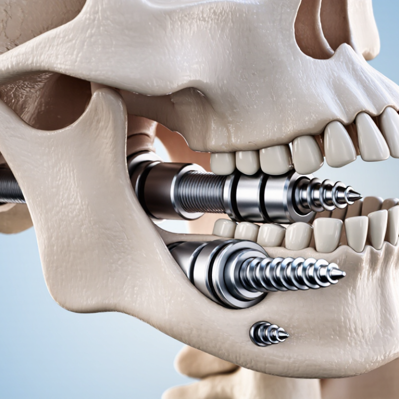
Cortical fixation implant systems are designed to secure orthopedic implants in dense cortical bone. Cortical bone forms the strong outer shell of many bones. It is thinner than cancellous bone, yet often provides reliable mechanical support. These systems use carefully shaped screws, plates, or fixation components to engage this outer layer. Their purpose is to improve stability during healing, movement, or load transfer.

In clinical practice, selection depends on bone quality, anatomy, injury pattern, and the patient’s daily demands. Imaging helps surgeons study cortical thickness before treatment. Surgical experience also matters, because inaccurate positioning may reduce fixation strength or irritate nearby tissue. Evidence supports cortical fixation in selected cases, but no system works equally well for every patient. That detail deserves attention. A technically advanced implant cannot replace careful planning, proper placement, and follow-up assessment.

Tips: Ask how the implant engages cortical bone. Discuss expected healing time, activity limits, and possible complications with a qualified orthopedic professional. Keep imaging records available when seeking another opinion. Small details can change the decision. A second review may reveal concerns that were missed during a busy consultation.

Why Choose Cortical Fixation Implant Systems?

Cortical fixation systems are designed to obtain screw purchase in the dense outer layer of bone. Compared with cancellous bone, cortical bone has substantially higher density and elastic modulus, which can improve mechanical anchorage and reduce reliance on the weaker internal bone structure. The chart shows representative midpoint ratios derived from commonly reported orthopedic biomechanics ranges; it is not a measurement of implant performance.

Reference ranges used for the calculation: cortical bone density approximately 1.8–2.0 g/cm³ and elastic modulus approximately 7–30 GPa; cancellous bone density approximately 0.1–0.3 g/cm³ and elastic modulus approximately 0.05–0.5 GPa.

How Cortical Fixation Supports Implant Stability

Why Choose Cortical Fixation Implant Systems?

How Cortical Fixation Supports Implant Stability

Cortical fixation supports implant stability by engaging dense outer bone. This bone is usually stronger than the softer interior structure. When fixation reaches reliable cortical purchase, the implant can resist unwanted movement during early healing. That matters because excessive micromotion may interfere with bone integration. The result is not instant success. It is a carefully managed starting point.

In clinical planning, surgeons assess bone quality, anatomy, loading demands, and the patient’s healing capacity. Cortical fixation may improve initial mechanical support when available bone is limited. It can also help distribute forces across a broader area. Small details matter, including insertion angle, preparation accuracy, and the implant’s contact with bone. Experience remains essential here. A strong concept can still perform poorly when planning is rushed.

Stability is not automatic.

Healing biology continues after surgery. Occlusal forces, hygiene, smoking, systemic conditions, and patient compliance may influence the outcome. For this reason, cortical fixation should be selected through individualized evaluation rather than treated as a universal solution. Clinicians should review imaging, confirm suitable anatomy, and explain realistic expectations. Even with careful technique, uncertainty remains. That honest recognition supports safer decisions and more reliable long-term care.

Key Design Features of Cortical Fixation Systems

Why Choose Cortical Fixation Implant Systems?

Key Design Features of Cortical Fixation Systems

Cortical fixation systems are designed to engage strong outer bone layers for controlled implant stability. Their screws often use fine threads, a compact pitch, and a narrower core. These features can improve purchase when available bone is limited. Thread depth matters. Too aggressive, and insertion may become difficult. Too shallow, and holding strength may suffer.

A reliable system should also support accurate alignment during placement. Locking interfaces can help maintain screw position, while a low-profile plate may reduce soft-tissue irritation. Some systems offer variable-angle control, allowing surgeons to adapt fixation to anatomy and fracture patterns. Material selection, surface finish, and imaging compatibility also influence clinical handling. In practice, small design differences become obvious under pressure. Instrument fit is one example. A theoretically excellent implant can still disappoint if the driver feels unstable or visibility is poor. Surgeon experience and patient anatomy remain decisive.

Tips: Review thread geometry, locking security, and instrument ergonomics before selection. Confirm that the system suits bone quality, loading demands, and imaging requirements. Do not treat “stronger” as universally better. Excessive stiffness may alter load distribution, while insufficient fixation may risk movement. Careful planning, appropriate sizing, and postoperative monitoring are essential. Even well-designed systems require thoughtful use.

Clinical Situations Where Cortical Fixation May Be Chosen

Cortical fixation may be chosen when metaphyseal bone cannot provide dependable support. This situation appears in osteoporotic, revision, and trauma cases. The International Osteoporosis Foundation reports that one in three women and one in five men over 50 may experience an osteoporotic fracture. Thin cancellous bone can collapse around an implant. Dense cortical bone may offer stronger purchase.

Surgeons may consider cortical fixation when immediate axial stability is important. Examples include poor bone quality, segmental bone loss, and limited implant length. It may also help when a conventional stem cannot achieve reliable metaphyseal contact. In practice, imaging, bone density, fracture pattern, and patient activity all influence the decision. Small details matter. A tight fit on a scan may still fail under repeated loading.

Large registry data provide useful context, but not perfect answers. The 2024 National Joint Registry report records more than four million hip and knee procedures, yet it does not separately measure every cortical fixation design. That limitation matters. Comparative studies often involve small groups, different surgical techniques, and short follow-up periods. A 2022 systematic review in the Journal of Orthopaedic Trauma also noted variation in fixation outcomes across patient populations. Cortical fixation should therefore be selected for a defined mechanical problem, not used automatically. Careful preparation remains essential. Even experienced teams can misjudge bone quality.

Why Choose Cortical Fixation Implant Systems? - Clinical Situations Where Cortical Fixation May Be Chosen

Clinical Situation Why Cortical Fixation May Be Considered Potential Clinical Benefit Important Considerations
Reduced bone quality
Including osteopenia or osteoporosis
The trajectory is designed to engage a greater proportion of dense cortical bone along the screw path rather than relying primarily on cancellous bone. May improve screw purchase in selected patients and may help reduce the risk of fixation loosening when appropriate technique and construct planning are used. Bone density, vertebral anatomy, fracture risk, and the need for augmentation should be assessed individually. Cortical fixation does not eliminate mechanical failure risk.
Short or narrow pedicles A cortical-based trajectory may provide an alternative path when conventional pedicle screw placement is limited by pedicle dimensions or morphology. Can support fixation planning in anatomically constrained segments while allowing the surgeon to select a trajectory suited to the patient’s anatomy. Thin-slice CT and multiplanar imaging are important. The technique requires precise entry-point selection and careful control of medial, lateral, cranial, and caudal angulation.
Lower lumbar fixation
Especially at the lumbosacral junction
The technique can be used to target cortical bone in the posterior elements and vertebral body, depending on the planned level and trajectory. May provide an alternative fixation strategy in patients where preservation of posterior anatomy or avoidance of a more lateral trajectory is clinically desirable. The construct must be evaluated for alignment, rod contour, interbody support, sacral anatomy, and the mechanical demands of the intended fusion.
Revision surgery
When previous fixation has altered the available anatomy
A different screw trajectory may help the surgeon work around a prior screw channel, provided that sufficient bone remains and the new path is safe. May expand fixation options and reduce reliance on the original trajectory in selected revision cases. Prior hardware, fusion mass, scar tissue, bone defects, and altered landmarks must be evaluated with current imaging. Navigation or intraoperative imaging may be appropriate.
Need to limit lateral muscle dissection The entry point is more medial than that used for many traditional pedicle screw techniques, which may support a less lateral exposure in suitable procedures. May reduce the extent of muscle dissection and support minimally invasive or muscle-sparing surgical approaches when clinically appropriate. Potential soft-tissue benefits depend on the complete surgical approach, incision placement, instrumentation, surgeon experience, and patient-specific factors.
Multilevel degenerative disease
When multiple levels require stabilization
Cortical fixation may offer an additional trajectory option for constructing a multilevel posterior fixation system while adapting to individual vertebral anatomy. Can contribute to a customized fixation plan and may be combined with decompression or interbody procedures when indicated. Global sagittal alignment, junctional stress, fusion levels, adjacent-segment condition, and overall construct stiffness should be considered.
Need for a less prominent posterior profile In selected cases, a medial entry point and shorter lateral excursion may support implant positioning closer to the midline. May be useful when posterior soft-tissue coverage or implant prominence is a concern, subject to the selected implant and surgical plan. Implant prominence is influenced by anatomy, rod and connector configuration, screw head design, body habitus, and postoperative positioning.
Cases requiring individualized fixation strategy
Based on anatomy, pathology, and surgical objectives
Cortical fixation can be selected as an alternative or complementary option when conventional trajectories do not provide the preferred balance of bone purchase, exposure, and construct design. Supports patient-specific planning rather than a one-trajectory-fits-all approach. Selection should be based on clinical examination, radiographic evaluation, pathology, bone quality, surgeon expertise, and the risks and benefits of available alternatives.
Clinical note: Cortical fixation is not universally superior to traditional fixation. Its suitability depends on patient anatomy, bone quality, pathology, imaging findings, surgical goals, and the surgeon’s training and judgment. This table is for educational use and does not replace clinical evaluation or surgical decision-making.

Factors for Comparing Cortical Fixation Implant Options

Why Choose Cortical Fixation Implant Systems?

Factors for Comparing Cortical Fixation Implant Options

Cortical fixation systems can provide strong purchase in dense outer bone. However, selecting one requires more than checking material or price. Surgeons should compare fixation strength, screw geometry, insertion control, and compatibility with the intended procedure. A well-designed system should support predictable placement, even when anatomy is narrow or bone quality varies. Small differences matter.

Clinical experience also highlights practical details. Does the implant offer clear size options? Can the surgeon adjust positioning without excessive force? Are the instruments ergonomic and easy to clean? Imaging visibility may influence postoperative assessment. Published evidence, regulatory documentation, and transparent testing deserve close review. Marketing language alone is not enough. I have seen specifications look impressive, yet the operating workflow felt less intuitive. That gap deserves attention.

Tips: Compare published biomechanical data with real clinical needs. Review complication reports and available training materials. Confirm that the system matches the patient’s anatomy, bone condition, and surgical plan. Ask whether insertion steps remain manageable under limited exposure. Also examine inventory demands, because missing sizes can interrupt a carefully planned procedure. No option is perfect. Careful comparison, surgeon judgment, and current clinical guidance should shape the final decision.

volume-unmute-yellow-icon volume-unmute-blue-icon